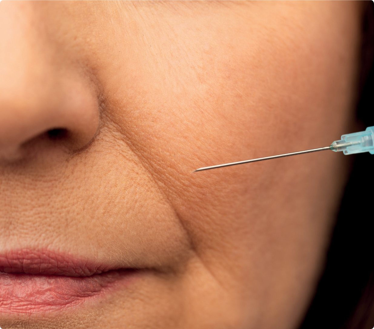
Hyaluronic acid dermal fillers are Food and Drug Administration (FDA)-approved for the correction of nasolabial folds through local dermal injections. In the past, dermal fillers were used to directly treat into the location of the desired correction. This would be through careful placement of the filler into the deep layer of the dermis and/or the subcutaneous tissue through a linear thread or fanning technique (Choi, 2015).
Despite good clinical results in the form of reduced appearances of wrinkles in the nasolabial fold, there are many pitfalls with the traditional technique. This ranges from something as simple as bruising to more sinister complications, such as vascular occlusion leading to alar necrosis and visual loss (Funt and Pavicic, 2013). This occurs due to the variability of the vasculature that runs at the subcutaneous level at the nasolabial fold (Hartstein, 2012). Furthermore, it became apparent that reduction of a nasolabial fold did not necessarily enhance the aesthetics of the patient, and in some cases treatment could look unnatural, owing to complete obliteration of the transition from the mid-cheek to perioral region. As our understanding of the facial aging process has improved, so too has our approach to treating the nasolabial fold.
Facial aging process
The process of facial aging is complex and is a cumulative effect of simultaneous changes of different facial tissues. It can be explained on an anatomical basis, as changes with aging occur at every level of the facial anatomy.
In general, the youthful face has the appearance of fullness and the aging face is characterised by sagging and volume loss. If we look at the skin, the process of aging is influenced by genetics, hormones, metabolic changes and environmental exposure. Oxidative stress is considered an important driver of the aging process with the production of reactive oxygen species (ROS), which causes connective tissue damage and decreased pro-collagen synthesis. Photodamage upregulates matrix metalloproteinase activity, further degrading dermal compounds. As the skin ages, it starts to become thin and flattened, with a gradual loss in elasticity. This is a result of decreased levels of fibroblasts and collagen (especially types 1 and 3) and elastin in the dermis. This ultimately leads to atrophy of the extracellular matrix and, with the integrity of the skin being compromised, the dermis loses the ability to resist the constant force of the muscles and lines start to appear in the skin, first during expression and eventually at rest (Mendelson, 2013).
The subcutaneous tissue consists of the fibrous and fat components, which are arranged in discrete compartments. The boundaries of the compartments correlate to the locations of the retaining ligaments and fibrous septa. The prominence of these subcutaneous fat pads over specific sites have been given specific names, such as the nasolabial fat. Aging causes concavities and convexities to develop between these compartments, making the transition between the compartments less smooth and more discernible. This is attributed to a number of causes, including selective atrophy and hypertrophy and attenuation of retaining ligaments, which causes malpositioning of the fat compartments. Unlike previously thought, fat minimally descends with aging (Mendelson, 2013). Studies have suggested that selective atrophy of deep fat compartments and relative hypertrophy of superficial fat compartments occurs with aging (Surek, 2016). This leads to larger size of adipocytes in superficial fat when compared with the deep fat (Surek, 2016).
It is widely known that the facial skeleton changes significantly with aging and that this has a profound impact on the appearance of the face. Peak skeletal projection is attained in early adulthood and, from this point, certain areas of the facial skeleton undergo dramatic resorption, while other areas continue to expand. Significant resorption occurs in the superomedial and inferolateral aspects of the orbital rim and the midface skeleton—in particular, the maxilla and the pyriform area of the nose, as well as the prejowl area of the mandible (Mendelson, 2013). The pyriform area recesses and the maxilla rotates with age, diminishing the anterior cheek projection and causing the deep medial cheek fat to fall laterally. Deficiencies in the skeletal foundation cause significant effects on the overlying soft tissue, such as the formation of the tear-trough or the nasolabial fold in the mid-cheek region (Surek, 2016).
Nasolabial fold anatomy
The terms ‘nasolabial fold’ and ‘nasolabial crease’ are used interchangeably. If we take a look at the international anatomic nomenclature, the nasolabial crease is a well-demarcated depressed facial line that occurs between the cheek and the upper lip from the ala of the nose to the corner of the mouth (Ramirez, 1995). On the other hand, the nasolabial fold is a bulge lateral to the crease—a ‘folding over’ of the fat pad and skin over a relatively fixed crease (Rudkin, 1999).
The nasolabial fold is made up of the following structures:
The nasolabial fold is a highly vascular area with the angular artery, arising from the facial artery, running in the subcutaneous fat of the nasolabial fold. The common understanding is that the facial artery becomes the angular artery after it gives off the superior labial artery. Looking at dissection findings, the angular artery is more likely to be within close relationship to the skin than the bone (Rubin, 1999). A 3-dimensional cranial computed tomographic scan shows that arteries are seen immediately below the skin at a distance of no greater than 4 mm (Cotofana, 2019). The deep pyriform space, a midface cavity, is bound medially by the depressor septi nasi and laterally and superficially by the deep medial cheek fat and lip elevators. Within this space, the angular artery courses at the roof of the space within a septum between the space and deep medial cheek fat (Surek, 2016).

Treatment planning
The below approach is the authors' recommendations, based on clinical experience.
The nasolabial fold must be addressed within context of how the aging process affects the face globally. Examination should begin with assessing the shape of the face, at rest and in dynamics. In the aging patient, volume loss can be examined in the mid face, which provides opportunity for revolumisation and indirect correction of the fold. The sequence in which the volume is lost give clues for the order in which to approach the mid face, to most effectively treat the nasolabial fold.
To address bony resorption, treatments along the zygoma can expand the superficial muscular aponeurotic system (SMAS) if placed supraperiosteally. SMAS expansion will benefit the nasolabial folds, as lateral tension is placed upon the tissue. Treatment deep at the pyriform fossa will address maxillary bony volume loss, and is an excellent treatment for a nasolabial fold, alongside supporting a drooping nasal tip, which also occurs with aging. The authors' recommend a high G’, elastic filler when treating supraperiosteally, to achieve maximal projection. Treatment to the pyriform fossa must be deep onto bone to avoid the angular artery.
To address deep fat volume loss, we must look to the deep medial cheek fat and the medial sub orbicularis oculi fat (SOOF). Restoring medial cheek volume within these fat pads can dramatically lift a heavy nasolabial fold owing to correction of migration and atrophy of these soft tissues. A cannula approach is recommended sub-SMAS and using a cohesive, high G’ filler to mimic the native deep fat tissue. Treatment can also be sited within the prezygomatic space adjunctive to the supraperiosteal zygoma treatment. This will further enhance cheek definition and further indirectly treat the nasolabial fold.
We can then address the superficial fat. It is important to avoid superficial fat where there is relative hypertrophy (infraorbital fat and nasolabial fat) and focus on areas where there is relative atrophy. If the patient has pre-auricular superficial fat volume loss, a good option is to use a cannula approach to fan a cohesive filler in the subcutaneous plane. This will create a lateral vector on the tissues and can soften nasolabial folds, and will particularly lift any early jowling too.
Finally, we can approach the nasolabial fold directly. This is best treated in the subdermal plane with a cannula, using a moderately elastic, cohesive filler to mimic the existing superficial fat. It is important to stay medial to the nasolabial fold itself, to avoid adding heaviness to the fold.
Through following an approach that addresses volume loss first, we can achieve complete correction of a nasolabial fold, often without needing direct treatment.
Case study
The patient was a 33-year-old female who attended clinic for full face treatment. On examination, she has balanced facial thirds and excellent symmetry.
Volume loss was noted at the:
The patient underwent a pan-facial filler treatment to rejuvenate the face, focusing on two main areas of concern: nasolabial folds and tired under-eyes.
Treatment plan
After careful consultation, the following treatment plan was agreed upon by the healthcare professional and the patient:
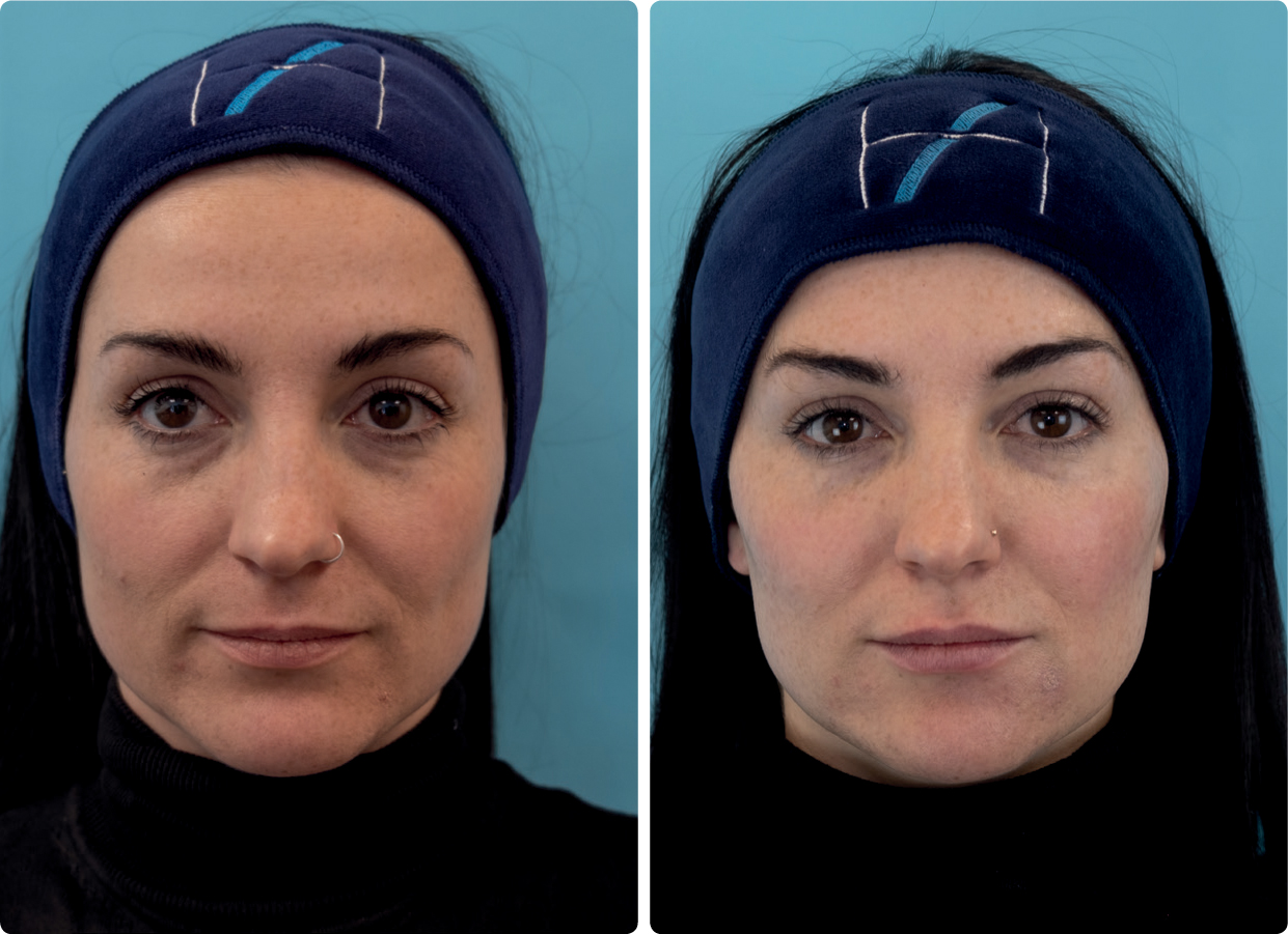
This treatment fully corrected the patient's nasolabial folds. Further treatment included toxin to three upper face areas and masseter muscles, alongside jawline and lip filler. No direct treatment was required to the nasolabial fold. Figure 1 shows the patient before and 4 weeks after treatment.

Conclusion
Today more than ever, patients have high expectations from their treatments and expect their practitioner to create treatment plans which are bespoke and effective.
We are entering an era in facial aesthetics where unhelpful old habits (such as treating lines directly rather than understanding their cause) are dying out. Practitioners must be equipped with an excellent understanding of the facial aging process and anatomy in order to achieve balanced natural results which truly address our patient's concerns and offer long lasting rejuvenation.
The mid face represents the foundation of the aging face, and correctly sequenced revolumisation of this region is an essential procedure for every aesthetic practitioner to master.


