


The re-positioning of tissues of the face is dependent on thread engagement in the superficial fatty tissue. The depth of this tissue varies in every face due to factors such as sex, age and sun exposure, etc.
Therefore, using one size of thread in the facial superficial muscular aponeurotic system (SMAS) and hoping to get optimum results cannot work. Surely the depth of the SMAS should determine the thread gauge (diameter)?
In clinical practice, this means that a smaller gauge thread should be used in areas of low fat depth, such as the pre-auricular and upper maxillary area. In the marionette area and the pre-jowl sulcus, a larger gauge (diameter) thread should be used.
To overcome this problem, in the past, the author pioneered the double twist technique. This technique can still be used with the new multi-diameter approach. It results in double vector force when used in either the direct or indirect route. It is not only capable of double vectorisation but, in certain circumstances, triple or even quadruple vectorisation has been used. The latter has mainly been used by the author in the male face.
It is known that caudal migration of the facial fat pads occurs, leading to facial tissue ptosis, especially in the lower two-thirds of the face (Wollina et al, 2017). There are three types of fat pads: superficial, deep and deepest. The third includes the buccal and temporal fat pads (Rohrich and Pessa, 2007). The size, position and associated ligaments of the superficial fat pads are relevant to this article.
» The area also has a delicate framework of the lymphatic drainage network, so too much manual manipulation should be avoided to preserve this structure «
In clinical practice, out of the three types of intra-adipose bands, the bands of the first order are important in the ability of the thread to achieve ‘tissue grab’ in the dermal white adipose tissue (WAT). Due to the rich, three-dimensional septal network in the medial, lateral as well as the parts of the periorbital region, temple and forehead, the adherence of the adipose tissue to the skin is loose. In clinical practice, this means that the re-positioning of the fat pads with threads in these areas will not take the skin with it to the same extent as other areas of the face. Therefore, relapse is more common.
In the malar and jowl areas, the SMAS is more distinct. Superiorly, the SMAS merges with the temporal fascia and, inferiorly, it merges with the platysma muscle.
The exact anatomy and distribution of the SMAS will not be discussed in detail here. Suffice it to say that the SMAS has three layers: superficial, middle and deep. The superficial layer is directly connected to the subcutaneous layer. This is the layer in which polydioxanone (PDO) threads are placed.
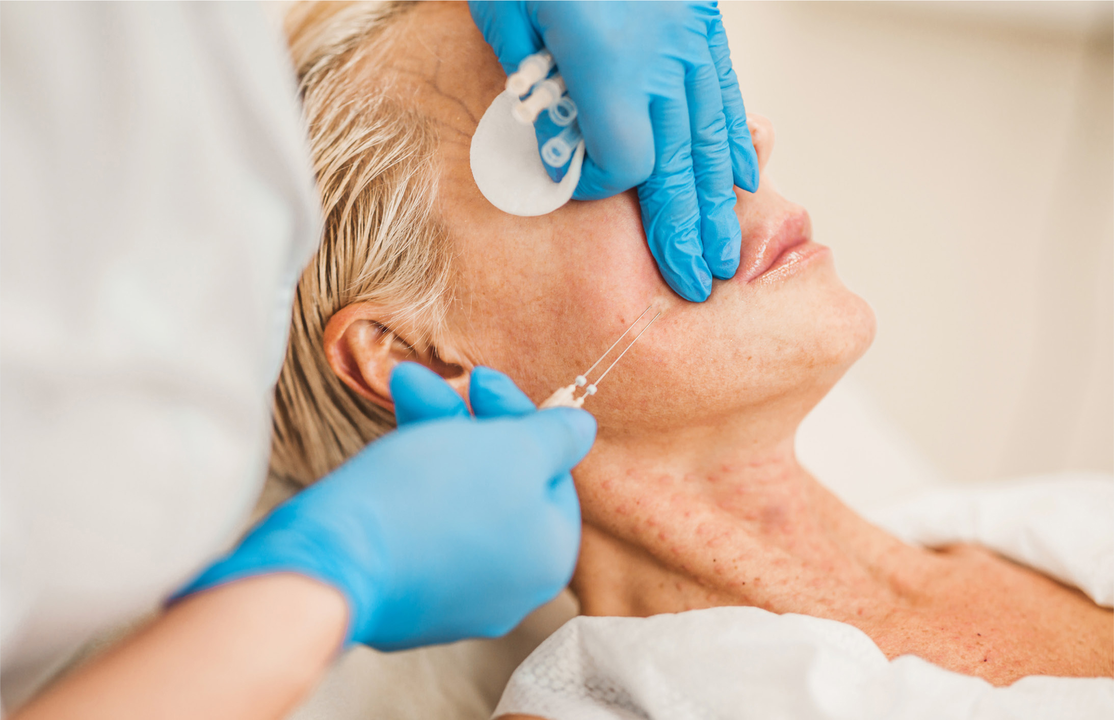
In clinical practice, the PDO threads are inserted at variable depths, from superficial to deep, starting from the zygoma to the lower face. The latter includes the nasolabial folds, marionette lines and pre-jowl sulcus.
Optimum results are achieved by further refining these procedures. Pre-positioning is useful and can be achieved by suture guidance or facial banding. Other techniques include gravitational positioning, manual manipulation, hydro-dissection, mechanical dissection, thread manipulation, hydroxo enhancement and combination techniques.
Thread manipulation is a technique developed by the author, which has been used in his clinical practice for the past few years. It can be used in several areas where thread procedures are carried out to enhance clinical outcomes. One area—the maxilla—will be described in some detail to achieve maxillary enhancement.
Maxillary enhancement
The cheeks have a large amount of superficial fat, which, by its loose arrangement, is very mobile. Therefore, the PDO threads placed in the cheeks give a good lift when inserted in the correct superficial musculoaponeurotic system plane. They can also be manipulated to achieve maxillary fullness without the need for other treatment modalities. This volumetric enhancement is achieved by an insert-and-push technique. It is an effective technique to use when there has been a loss of elastin and collagen and thinning of the mimetic muscles.
The technique must be correctly carried out, and the author recommends thorough training before it is implemented. If this is not the case, the reader is warned that damage to the infraorbital nerve, vein and even the artery can occur. The area also has a delicate framework of the lymphatic drainage network, so too much manual manipulation should be avoided to preserve this structure.
Clinical technique
There is no substitute for proper hands-on training and certification, and the following description is for educational purposes only.
With the patient seated in the upright position, the infraorbital foramen is identified and outlined with a skin pencil. A circle is drawn with a diameter of 1.5cm with the infraorbital foramen being in its centre. It is very important that no manual manipulation is performed in this area.
From the entry point, the thread is inserted in the usual way to the vertically dropped lateral canthus line. Keeping the cannula slightly raised, an insert and upward push manoeuvre is performed, advancing 1cm at a time to 0.5cm past the mid-pupillary line. The cannula is then slowly removed with a sweeping manoeuvre, ensuring that the notch in the ‘W’ type cannula is facing towards the deeper tissues
When correctly performed, the technique will raise the malar fat pad, which lies superficial to the SMAS and directly below the lateral part of the infraorbital rim. This pad provides the bulk of the convexity associated with a youthful appearance, so its enhancement is beneficial. The malar fat pad is about 6mm thick and overlies the maxilla. The subdivision of this pad is done based on anatomical partitions but, for the clinician's purpose, this subdivision is irrelevant. The pad is loosely attached to the overlying dermis, along with the lateral orbital thickening and the orbicularis retaining ligament. These hold the pad in the superolateral position, albeit loosely. It is important to note that no connective tissue attachments have ever been demonstrated to the SMAS under the pad. This makes the malar fat pad prone to gravitational forces, in addition to ligamentous laxity resulting from ageing. This fact is also used to achieve maxillary enhancement using the technique described above using the PDO thread to achieve cephalic re-positioning.